Researchers at the University of Surrey developed an AI that predicts future knee X-rays, helping doctors and patients visualize osteoarthritis progression with greater accuracy, speed, and transparency for earlier, personalized care.
A revolutionary artificial intelligence system created by researchers at the University of Surrey is redefining how doctors and patients can visualize and predict arthritis progression. The AI can forecast what a patient’s knee X-ray will look like a year into the future, offering both a visual projection and a personalized risk score that together could transform osteoarthritis care.
Seeing the Future of Osteoarthritis
Osteoarthritis affects more than 500 million people worldwide, making it the most common form of arthritis and a leading cause of disability among older adults. Until now, tracking its progression relied on periodic X-rays and subjective assessments — often leaving patients in uncertainty about how their condition might evolve.
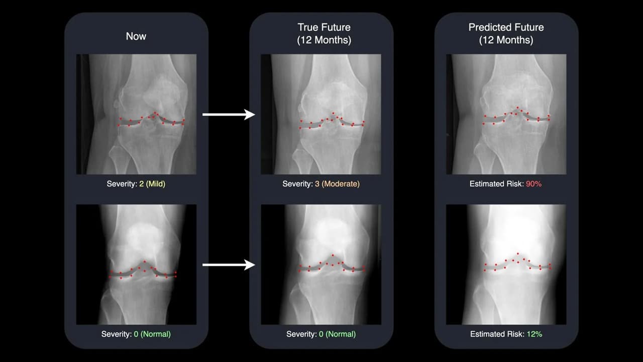
The Surrey AI changes that. By analyzing thousands of knee images, it generates a realistic “future” X-ray, showing doctors and patients exactly how a joint could change over the coming year. The system also provides a numerical risk score, helping clinicians identify patients most likely to experience rapid deterioration.
This innovation was presented at the International Conference on Medical Image Computing and Computer Assisted Intervention (MICCAI 2025) and marks a major leap in the use of AI for personalized, predictive medicine.
Faster, Clearer, and More Human
The model was trained on nearly 50,000 X-rays from 5,000 patients, one of the largest datasets of its kind. It can predict changes up to nine times faster than previous AI systems, while also delivering more interpretable results.
Lead researcher David Butler from Surrey’s Centre for Vision, Speech and Signal Processing (CVSSP) explained:
“Most AI tools give a number or a simple prediction, but not much explanation. Ours actually shows you what your future knee could look like. Seeing your current and future X-rays side by side can be incredibly motivating — it helps doctors intervene earlier and encourages patients to stay on track with their treatment or lifestyle changes.”
This human-centered approach bridges the gap between complex algorithms and real-world healthcare, offering clarity where uncertainty once existed.
How the AI Works
At its core, the system uses a diffusion model, a type of generative AI that creates realistic images by learning patterns of change over time. It tracks 16 specific points within the knee joint, mapping how cartilage, bone spacing, and other features might evolve.
Unlike traditional black-box algorithms, this model offers transparency — clinicians can see exactly which parts of the knee the AI is monitoring and why. That interpretability builds trust, making the system easier to adopt in hospitals and clinics.
A Glimpse Into the Future of Medicine
The Surrey research team believes this approach could extend beyond arthritis. Similar systems might one day predict lung damage in smokers, track heart disease progression, or even visualize tumor growth patterns, offering doctors a visual timeline of disease development.
Professor Gustavo Carneiro, a senior researcher on the project, emphasized the broader potential:
“Earlier AI models could calculate risk, but they couldn’t show it. Our tool combines speed, transparency, and visualization — helping doctors spot high-risk patients earlier and personalize their care in ways that weren’t possible before.”
Why It Matters
For millions living with osteoarthritis, the fear of worsening pain or losing mobility can be overwhelming. This technology offers something new — a glimpse of the future that empowers patients and clinicians alike. By visualizing progression before it happens, it opens the door to timely treatment, lifestyle changes, and hope for better outcomes.